Have you had excessive radiation exposure at the dentist?
When you go to the dentist you should have x-ray images taken of your teeth every 12 months. Radiographs and other imaging methods are used to check for oral diseases, as well as to monitor dentofacial development. However, the dentist must weigh the benefits of taking dental radiographs against the risk of exposing a patient to x-rays, the effects of which accumulate from multiple sources over time. So how much radiation are you exposed to when at the dental office? The answer may surprise you.
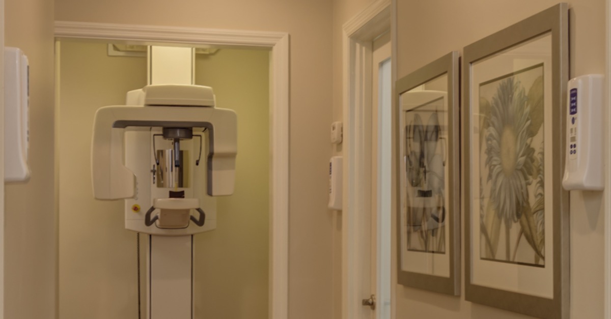
Radiographic examinations can be performed using digital imaging or conventional film. Digital imaging may offer reduced radiation exposure and the advantage of image analysis that may enhance sensitivity and reduce error introduced by subjective analysis. When you see your dentist at a cleaning they will probably take a few x-ray images. The exposure rate for a full mouth series of x-ray images (patients rarely need this many x-rays) is equal to only three millirems. A millirem is fancy radiation talk for a dose of radiation. Three millirems are equivalent to about four days of exposure to radiation received naturally from the sun. A panoramic x-ray is made by the big machine that spins around your head. It provides images of your sinus cavity, jaw bone, as well as your teeth. The radiation exposure from this imaging device is close to what you would get on a one way plane ride from Knoxville to Denver.
ALARA is an acronym for As Low As Reasonably Achievable. This is a radiation safety principle for reducing radiation doses by employing all reasonable methods. Even though dental x-rays expose the patient to a very low dose of radiation, we still try to limit the exposure. Here is the criteria that our Knoxville dental office uses to prescribe dental x-rays.
- Children – Children generally need more X-rays than adults because their teeth and jaws are still developing and because their teeth are smaller. As a result, decay can reach the inner part of the tooth, dentin, quicker and spread faster. We also monitor the child’s erupting teeth in case early intervention orthodontics are needed.
- Adults with extensive restorative work, such as fillings – We look for decay beneath existing fillings and crowns or in new locations.
- People who drink a lot of sugary beverages – We look for tooth decay due to the high sugar environment. Sugar creates a perfect situation for cavities to develop.
- People with periodontal (gum) disease – It is critical to monitor bone loss in patients with gum disease. Progressive bone loss will eventually lead to tooth loss.
- People who have dry mouth – Dry mouth is called xerostomia. It can be caused by taking several medications, certain disease states like Sjogren’s syndrome and cancer treatments. Constant dry mouth can lead to the development of cavities.
- Smokers – Smokers are at increased risk of periodontal disease.
Our Knoxville dental office is the first in the East Tennessee area to implement the new Planmeca ProSensor HD, a digital x-ray sensor that creates the most clear and crisp image while using low levels of radiation. This state-of-the-art technology allows us to make an accurate diagnosis while ensuring that our patients get the best possible care though reduced radiation levels. All of our dental assistants are certified in the use of radiography. We want you to feel completely comfortable in our office and encourage you to discuss and radiation concerns with our dental team. Remember, you may receive more radiation popping pop corn in the microwave than you receive from one of our X-rays.
Related Posts